|
|
|
| Select a topic: |
Heart Registry | Tricuspid Valve Dysplasia and Subaortic Stenosis |
| Samoyed State Of Heart |
|
|||||
Samoyed State of Heart
by Cheri Hollenback |
|||||
|
Over the ages, the heart has come to symbolize many qualities. Courage, stamina, “showability” and the quality of an affectionate temperament have all been described as having “heart”. Although we see our Sammies consistently demonstrating these qualities, their anatomical “hearts” may also carry a less desired property -- that of congenital heart disease. In a review of 22,283 dogs conducted at the University of Pennsylvania during the years between 1987-1889, congenital heart disease (CHD) was found in 150 animals (0.67%). The Veterinary Medical Data Base, maintained at Purdue University, found the rate to be 0.85% in 154,233 dogs during the same time period (Buchanan, 1992). There is some concern in the Veterinary community that the rate of CHD may be increasing. However, it is unclear whether the actual frequency is greater or improved diagnosis and broader availability of veterinary cardiologists is raising the number of dogs diagnosed with CHD.
|
|||||
|
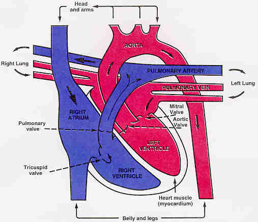
Considering these rates, of the 3199 Samoyeds registered with the American Kennel Club in 1997, we would anticipate a minimum of 21 to 27 of these puppies to have some type of CHD (AKC, 1998). Unfortunately, Samoyeds have higher rates of occurrence for two of the top three types of CHD. Based on the analysis performed on the 1320 dogs identified as having CHD in the aforementioned Veterinary Medical Data Base (VMDB), Samoyeds have a 2.8 fold greater risk of having aortic stenosis. Additionally, our Sammies have a 5.4 fold greater risk of pulmonic stenosis. When these factors are applied to the number of Samoyeds registered in 1997, the number of puppies we would predict to have aortic stenosis jumps to 59-76, while the number having pulmonic stenosis climbs between 113 to 146. These statistics become moot in the event your puppy has CHD. Suddenly, the statistic for your puppy -- and you -- is 100%. This is the situation I found myself in earlier this year when a puppy I had bred had the diagnosis of pulmonic stenosis confirmed. To understand CHD in Samoyeds, it is helpful to first understand some basic anatomy of the heart (image 1). The heart is mainly muscle tissue, but also contains one way valves, nerves, fibrous tissue and it’s own blood vessels which supply the heart with nutrients. The upper chambers are called the “atria” (plural) or “atrium” (singular), while the lower chambers are the “ventricles”. The heart’s four chambers provide two pumping circuits -- Pulmonic (right atria and ventricle) , and Systemic (left atria and ventricle). The pulmonic circuit has the easier task of circulating the blood the relatively short distance to the lungs and back to the left atria. The systemic circuit, particularly the left ventricle, works considerably harder, in that it must circulate the blood out to the entire body. After the blood passes through the capillaries, the series of one-way valves in the veins and the contractions of the muscles help return the blood to the heart. The systems working together can be envisioned as a figure eight laying on its side -- or the Greek symbol for infinity, with the heart sitting at the point of intersection. [TOP] |
|||||
|
NORMAL HEART
|
|||||
|
A freshly oxygenated red blood cell leaves the lungs by way of the pulmonary veins (PV). These veins -- who break the rule of veins carrying un-oxygenated blood -- empty into the left atrium (LA). The red blood cell then flows out of the atrium into the left ventricle (LV) through the mitral valve (MV). The left ventricle contracts, causing the flaps of the mitral valve to close, thus preventing blood from flowing backward (regurgitating) into the atrium. Additionally, the contraction ejects the oxygenated blood out through the aortic valve (AV) into the aorta. From there, the oxygenated blood flows to all parts of the body to provide the essential oxygen and other nutrients to the cells. After the cells are spent of their nutrients and pick up any waste products, the blood flows through the veins to the vena cava and back to the right atrium (RA) of the heart. It then flows through the tricuspid valve (TV) into the right ventricle (RV) which ejects the blood through the pulmonary valve (PV) back to the lungs via the pulmonary artery (PA). There, in the tiny capillaries of the lungs, the waste products such as carbon dioxide are removed and oxygen is picked up for the next go around. In the fetal puppy, there are a couple of shortcuts in or near the heart that bypass the lungs. Since the lungs do not provide any nutrients nor remove any waste products in the fetus, there is no reason for all the blood to go to the lungs. Just a small portion flows to the lungs to provide nourishment for the growing tissue. One of these shunts is a structure called the Ductus Arteriosis (DA). This little vessel creates a connection between the pulmonary artery and the aorta. By this shortcut, the blood leaving the right ventricle is allowed to go immediately to the aorta and return more quickly to the nutrients provided by the placenta. The other shortcut is in the wall between the right and left atria. There is a small opening in the tissues that allows the blood to flow from the right side to the left, again getting the blood back to the nutrient source more efficiently. These two shortcuts -- or shunts -- are usually open at the time of birth, but generally close over the first few hours to days as the lungs start performing their important role. Often if you listen with a stethoscope to the newborn puppy’s heart, you will be able to hear a murmur because of these shunts. |
|||||
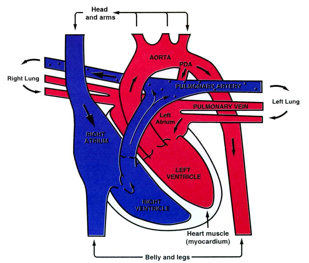
| The most common form of CHD found in dogs is Patent Ductus Arteriosis (PDA). In this condition, the ductus arteriosis does not close as it should so a significant amount of blood is able to flow “backward” across the ductus. This “leak” causes the left ventricle to have to work harder to circulate the blood to the systemic circulatory system. Subsequently, one of the symptoms is a bluish cast to the normally pink mucous membranes or cyanosis. Another symptom is a persistent heart murmur. Murmurs occur any time the flow of the blood is turbulent or the speed (velocity) of the blood flow increases. This phenomena is somewhat similar to the sound of water running through a hose changing pitch when the hose is pinched or twisted. The usual treatment for a PDA is to surgically close the ductus by tying it off (this is termed surgical ligation). A newer therapy is being explored whereby a thin catheter is inserted into a major vessel and a small coil is guided into the ductus to block it off. Fortunately, researchers have not found Samoyeds to be at increased risk for this particular problem. | |||||
|
PATENT DUCTUS ARTERIOSIS
|
|||||
|
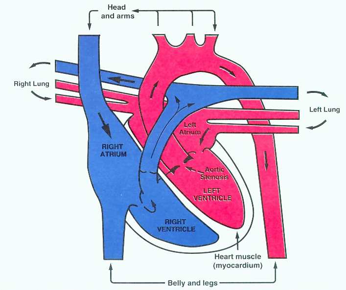
The second most commonly found heart defect reported from the VMDB is Aortic Stenosis (AS). In this condition, there may be three different anatomical defects that contribute to the narrowing of the outflow tract from the left ventricle. The most easily remedied problem is when the leaflets of the aortic valve are fused together, creating a narrow opening through which the blood may flow. Other presentations occur when the tissue at the base of the aorta or the base of the valve are narrow and fibrous, with the latter manifestation being the most common. The defect may also manifest with any combination of these presentations. The heart has to work extra hard to pump the blood against the restricted valve, causing the muscles of the ventricle to hypertrophy (enlarge). Aortic stenosis may be suspected in a puppy who has a persistent murmur, but without careful listening by a skilled veterinarian, it may remain undetected. As a youngster, the puppy’s heart will work to compensate for the narrowed valve, but over time, other symptoms -- including cyanosis (blueness to normally pink mucous membranes), exercise intolerance, congestive heart failure and sudden death, may occur. Twenty percent of dogs diagnosed with AS die unexpectedly, while with severe aortic stenosis, 70% of the dogs will die within the first three years of life. Treatment for this defect is dependent upon the anatomical structure of the defect, but generally, open heart surgery is the recommended treatment of choice. |
|||||
|
AORTIC STENOSIS
|
|||||
|
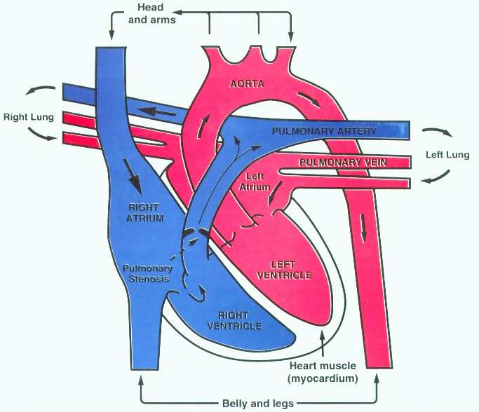
The third most common condition reported in all breeds of dogs is the defect most frequently seen in Samoyeds -- that of Pulmonic Stenosis. This defect is similar to AS in manifesting either as fused valve leaflets (valvular stenosis), narrowing in the tissue at the base of the valve (subvalvular) or the base of the pulmonic artery, or any combination of these. However, the condition is somewhat less ominous than AS, given that the defect is on the pulmonic side of the heart. Generally, the dog’s heart can compensate for the lesion as a youngster, but may develop more problem as it grows older. |
|||||
| PULMONIC STENOSIS
|
|||||
|
Diagnosis is first suspected when the dog has a persistent systolic murmur (present during the contraction phase -- or “lub” of the “lub-dub” of the heart beat). The diagnosis is confirmed with an EKG, X Rays of the heart, echocardiogram and cardiac catheterization (placing a flexible, narrow tube into a vessel and threading it to the area of the heart while watching the process on a fluoroscope. Often dye is injected to watch the flow of the blood.). The severity of the problem is determined by measuring the velocity of the blood flow across the narrowing and then calculating the pressure the heart is having to generate. Normal or near normal function of the heart can be restored in 70% of the affected dogs by performing a balloon valvuloplasty. In this procedure, a thin plastic catheter (tube) with a sausage shaped balloon on its end is passed through the narrowing. When in position, the balloon is inflated, stretching out the narrowed opening and rupturing any fusing between the valve leafs. Breeding experiments to determine the heritability of congenital heart disease supported the theory that the pattern of inheritance is polygenetic in nature (Buchanan, 1992). Veterinary Cardiologists strongly recommend that affected dogs are withdrawn from any breeding program. Non-affected siblings may be used with careful screening of mates and offspring. Non-affected animals who produce the defect in breeding with more than one bloodline are also recommended to be withdrawn from breeding programs (Thomas, 1998). Further research on the canine genome may allow more specific determinations of genetic risk for CHD in the future. |
|||||
|
Recommendations for affected animals are based on full identification of the extent and severity of the lesion. In the most severe of cases, particularly with Aortic Stenosis, immediate euthanasia is often the course of action. In less severely affected animals, surgical intervention, medications and other supportive treatments may provide improvement adequate to allow the animal a full life expectancy. The breeder’s choice of treatment options will also be impacted by personal philosophy, available resources and experience. Personally, I found myself in a dilemma when the diagnosis of Pulmonary Stenosis was made. My “head” was clear that my best course of action would be to have the dog euthanasized, but my “heart” could not support that choice for a puppy who was exhibiting no symptoms of the defect other than a murmur. After confirming the diagnosis with EKG, X-Ray, echocardiogram and cardiac catheterization at Washington State University, I flew the puppy to UC Davis where a balloon valvuloplasty was performed. That procedure was not as successful as hoped, due to a sub-valvular fibrotic ring. However, in July we returned to WSU for a follow-up examination and found that the procedure had adequate effect to allow the heart muscle to relax enough to decrease its size. The echocardiogram results indicated that the velocity of the blood through the defect had halved, allowing the pup to be reclassified as “mildly to moderately affected” rather than the previous diagnosis of “severely affected”. This improves his long term prognosis to have no effect on his anticipated lifespan. It was a joyful occasion to bring him home to his owner and share this wonderful news. “Lucky” - so dubbed by his owner - will return to WSU in one to two years for another evaluation. Although this is not necessary for his health, I feel it is important that he stay connected to the veterinary health system to provide any data regarding the long term prognosis of this defect. For that purpose, I have remained his co-owner. When he does make his journey to the Rainbow Bridge, his heart will be sent to UC Davis for study. Until that time, I expect he’ll continue jogging Lake Coeur d’Alene with his person, playing in his favorite pond and charming all with whom he comes in contact. We both owe a huge debt of gratitude to the Veterinary Medical professionals we have encountered on this trip, particularly Dr. Anthony Tobias of Washington State University (Go Cougars!) and Dr. William Thomas of UC Davis. |
|||||
| Glossary | |||||
|
|
|||||
References
|
|||||
| Images:
The heart images are provided, with permission, courtesy of Dr. Joel Hardin, MDPB, Department of Pediatrics, Southern Illinois University School of Medicine. These are images and descriptions of the human heart--the basic structures of the canine heart are similar, but the atria are smaller. Suggested Links:
|
|||||
| Last updated: Saturday, February 06, 2010 | |||||
 |
| | mirage home | health home | index | eyes | kidney | skin | orthopedic | | epilepsy | heart | endocrine | bloat | tick faq | search | health links | |